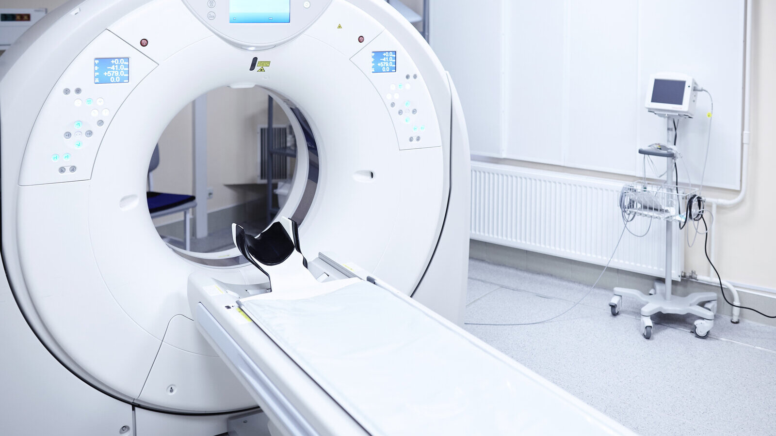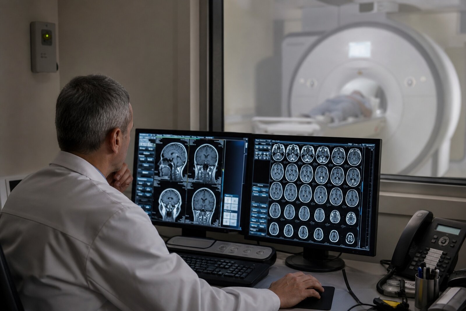

In November 2007, two radiation scientists, David Brenner and Eric Hall, wrote a review in the New England Journal of Medicine [1]. They estimated that more than 62 million CT scans were done each year in the United States. In 1980, the number was about 3 million.
A CT (computed tomography) scan takes X-ray pictures from many angles and builds them into 3D views of the body. Brenner and Hall warned that CT gives much larger doses than regular X-rays. They also wrote, plainly, that “the risks for any one person are not large.”
People still ask how much radiation a scan really gives, and the honest answer is a range rather than a single number. I think most people need a rough sense of the scale, an honest note about what is uncertain, and good questions to ask.
What does a millisievert measure?
When X-rays pass through your body, some of the energy is absorbed. That absorbed energy is what counts toward your radiation dose. RadiologyInfo.org, a patient website from radiology societies, explains the usual unit [2]. It is the millisievert (mSv). It measures effective dose, a number that estimates the harm from radiation to the whole body.
Effective dose also takes into account that some body tissues are more sensitive to radiation than others. If an exam “includes tissues or organs that are more sensitive to radiation,” the guide says, the effective dose will be higher. Because of this, very different kinds of radiation can be measured on one scale. That lets us compare a scan with the natural radiation around us.
This scale is useful, but it is only a rough guide. Brenner and Hall noted that it “provides only an approximate estimate of the true risk” [1]. To judge risk, they preferred to look at the dose each organ receives.
How does a CT scan compare with an X-ray?
The guide says the average American gets about 3 mSv per year from natural sources [2]. The largest part, about 2 mSv, comes from radon gas in homes.
One adult chest X-ray is about 0.1 mSv, or about 10 days of natural radiation. CT doses are larger. Here are the guide’s typical values for an average-sized adult.
A CT of the brain is about 1.6 mSv, similar to 7 months of natural radiation. A low-dose CT for lung cancer screening is about 1.5 mSv, or about 6 months. A CT of the chest is about 6.1 mSv, similar to 2 years. A CT of the abdomen and pelvis is about 7.7 mSv, or about 2.6 years.
If you divide 6.1 by 0.1, you find that a typical chest CT gives about as much radiation as 60 chest X-rays. A 2008 dose catalog by Mettler and colleagues lists average CT doses of about 2 to 20 mSv [3].
Why is there no single CT dose?
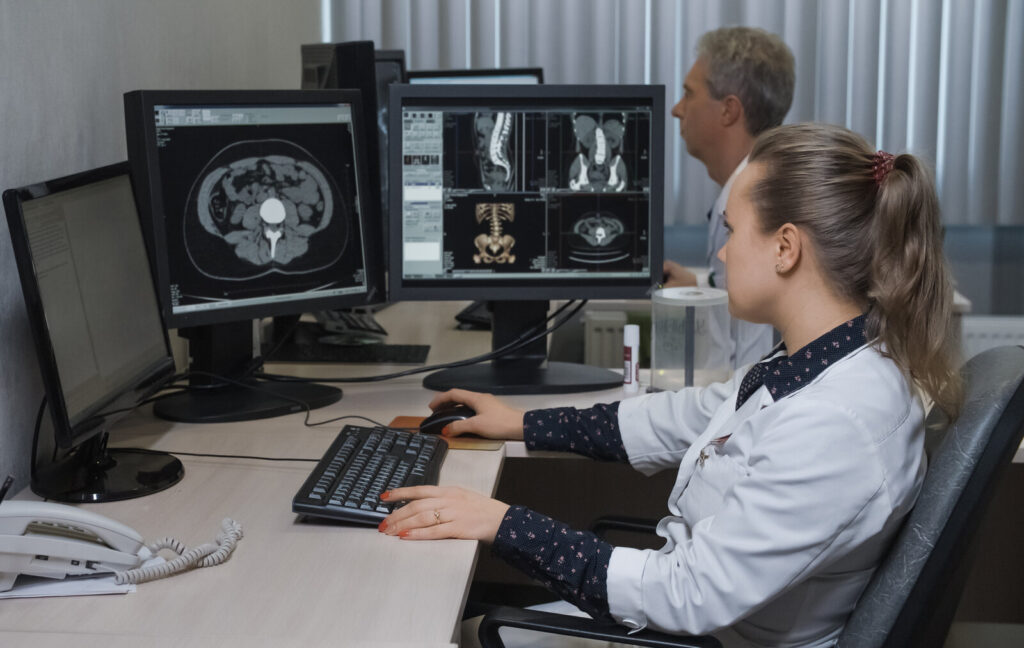
Those numbers are typical values. Your own dose may be different. The guide says the actual dose “can vary substantially, depending on a person’s size, the reason for imaging, and differences in imaging practices” [2].
An abdomen and pelvis CT done twice, with and without contrast dye, is about 15.4 mSv. That is double the single scan. Brenner and Hall listed other things that change the dose [1]. They include the machine settings, how much of the body is scanned and the scanner’s design.
The imaging team controls many of these settings. Brenner and Hall wrote that adjusting the settings to each patient’s size was “increasing but is by no means universal.” Lower doses also make grainier images that are harder to read. So the lowest dose is not always the best choice.
Imagine two coworkers who each had a CT last month. One had a quick scan of the head. The other had the abdomen and pelvis scanned twice, with and without dye. Both tests were called a CT. But one coworker received several times as much radiation as the other.
So when people ask how much radiation a CT gives, the honest answer depends on three things. It depends on which part of the body is scanned, on the person’s size, and on how the scan is done.
What is the cancer risk, and how sure are we?
At these doses, the main concern is a small added chance of cancer later in life. Brenner and Hall drew much of their evidence from survivors of the 1945 atomic bombs in Japan [1]. Survivors who received low doses, from 5 to 150 mSv, had a clear rise in cancer risk. These were doses to body organs. They are not measured the same way as the scan values above. Children face greater risk from the same dose. Their bodies are more sensitive to radiation, and they have more years of life in which a cancer could develop.
Brenner and Hall also made a rough estimate for the whole country. Based on how often CT was used in the US at that time, they suggested that perhaps 1.5% to 2.0% of all US cancers might be caused by CT radiation. That is a rough projection for the US population of 2007, not anyone’s personal risk.
Other experts read the evidence more cautiously. In 2023, the American Association of Physicists in Medicine (AAPM) published a position statement [4]. It accepts that in theory any dose could have some effect. But for most imaging tests, it finds “no conclusive epidemiological evidence” of harm. Epidemiological studies track health across large groups of people. The AAPM adds that risk predictions “are subject to significant uncertainty and should not unduly influence the decision for a justified procedure.”
In my view, these two groups of experts agree more than it first seems. Both treat the risk to one person from one needed scan as small. They differ on how sure we can be about the effect of small doses across millions of people. For a patient, the lesson is the same. No one can turn your scan’s dose in millisieverts into an exact personal cancer risk.
Questions to ask before a CT scan
Not having a needed scan also carries a risk: the doctor may miss an illness. The guide says “a person is at risk if the doctor cannot accurately diagnose an illness or injury” [2]. Brenner and Hall agreed that when a scan is medically needed, “the associated risk is small relative to the diagnostic information obtained” [1].
Brenner and Hall were most worried about scans that were not needed. An informal poll of pediatric radiologists, which they cited, suggested that perhaps one third of CT studies could be replaced or skipped. In some cases, a test without X-rays can be used instead, such as ultrasound or MRI (magnetic resonance imaging). They also noted that needed scans were sometimes repeated as patients moved from one doctor or hospital to another, often “simply because of a lack of communication.”
I am a researcher trained in medical physics. The scans that worry me most are the ones that were not needed. If a CT is suggested, these questions are fair to ask:
- How will this scan change my care?
- Have I had a recent scan that could answer the same question?
- Would ultrasound, MRI or a lower-dose scan work instead?
- For a child, will the settings be adjusted for their size?
These questions are not a reason to refuse a scan your doctor recommends. When there is a clear reason for a scan, its benefit is usually larger than its risk. Asking these questions helps make sure each scan is the right one.
In my view, a CT scan is not completely harmless, but it is also not something to fear. I would like people to know the rough size of the dose and accept that the risk estimate is uncertain. I think worry is best saved for scans that are not needed.
The numbers in this article are typical values, and they have limits. Your own dose depends on your body, the machine and the reason for the scan. Ask the imaging team about your exam, talk it over with your doctor, and decide together.
References
[1] D. J. Brenner and E. J. Hall, “Computed tomography: An increasing source of radiation exposure,” The New England Journal of Medicine, vol. 357, no. 22, pp. 2277-2284, Nov. 2007, doi: 10.1056/NEJMra072149.
[2] RadiologyInfo.org (Radiological Society of North America and American College of Radiology), “Radiation dose,” last reviewed Apr. 15, 2025. [Online]. Available: https://www.radiologyinfo.org/en/info/safety-xray
[3] F. A. Mettler, W. Huda, T. T. Yoshizumi, and M. Mahesh, “Effective doses in radiology and diagnostic nuclear medicine: A catalog,” Radiology, vol. 248, no. 1, pp. 254-263, Jul. 2008, doi: 10.1148/radiol.2481071451.
[4] American Association of Physicists in Medicine, “AAPM position statement on radiation risks from medical imaging procedures,” Policy PS 4-B, Nov. 16, 2023. [Online]. Available: https://www.aapm.org/org/policies/details.asp?id=318&type=PP¤t=true
Related Articles